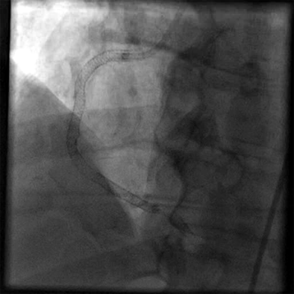

Occasional ventricular arrhythmias and even sudden cardiac death have been reported.įigure 1: Measurement of coronary flow by CTFC.
TIMI FLOW GRADE PRO
III - Pro gnosisĬoronary slow-flow phenomenon usually has a benign long term outcome but may be associated with relapses. Nitric oxide potentiating β-blocker nebivolol improved flow mediated dilatation of brachial artery and controlled chest pain in a study conducted by Albayrak et al ( 10). Statins with their pleiotropic effects on the vascular function have shown benefit in the treatment of CSFP ( 9). In an observational study, Kurtoglu et al found dypiridamole to be useful in the management of CSFP, with symptomatic as well as angiographic improvement ( 8). The data on conventional L-type calcium channel blockers use is scarce. However, due to its many drug to drug interactions, mibefradil is currently not in use. The T -type calcium channel blocker mibefradil was evaluated and was found to improve both clinical as well as angiographic outcomes ( 7). Treatment modalities for CSFP are not well established. There are case reports of abnormal QT dispersion and ventricular arrhythmias ( 6).Īffected patients are usually young male smokers. A significant proportion of them present to the emergency department with features of an acute coronary syndrome ( 5) and may have troponin elevations. Patients with CSFP often present with chest pain and ECG changes. The later is based upon images acquired at 30 frames/second and a correction factor of 1.7 for the LAD ( 4). TIMI-2 flow grade (ie requiring ≥ 3 beats to opacify the vessel) or a corrected TIMI frame count > 27 frames have been frequently used. The diagnosis of the CSFP can be made on the basis of the TIMI flow grade or TIMI frame count ( 3). Functional and morphological abnormalities in the microvasculature, endothelial dysfunction, raised inflammatory markers, occult atherosclerosis and anatomical factors of epicardial arteries have all been implicated ( 2). The exact pathogenesis of CSFP is still not clear and is probably multi factorial. Incidence of coronary slow-flow is reported to be 1-7% of all coronary angiograms. Moreover, the clinical profile and presentation of CSFP differs from syndrome X and is also different from coronary slow flow secondary to coronary ectasia or spasm, ventricular dysfunction, valvular heart disease and connective tissue disorders. Coronary slow-flow phenomenon was identified as an exclusive clinical entity in 1972 ( 1) where the distal opacification of the coronary artery is delayed on angiography in the absence of significant coronary artery disease.Ĭoronary slow flow phenomenon needs to be differentiated from slow-flow resulting from percutaneous coronary intervention.


 0 kommentar(er)
